

Practice Perfect 748
Stop Using the First Ray Excursion Test
Stop Using the First Ray Excursion Test

I’m on a crusade!
“A crusade?” you ask. “Are you trying to end hunger or achieve peace in the world?”
No, my friends, I’m on a crusade for podiatrists to stop using the first ray excursion test! Grandiose, right? Shapiro’s swinging for the fences on this one. This is going to make world history and change life as we know it. Are you already convinced?
Wait…
Do I hear a dissenter? “What the heck are you talking about, Shapiro? Are you nuts?” Well…yes, of course I’m crazy. We’re well aware of that. In this case, though, I’m crazy hating on this examination maneuver for the reason that it’s a liar. Yes, good colleagues, you’ve been fed a falsehood all these years that the first ray excursion test tells you that the first ray is “hypermobile” (whatever hypermobile means). We’ve all been taught that this test reveals excessive motion at the first tarsometatarsal joint. We’ve also been taught that this is a major indication for the Lapidus procedure in patients with bunions. I hear students and residents dutifully repeating this mantra in the hallowed hallways of our universities, clinics, and operating rooms.
WRONG!!!
Instead, there’s solid research to confidently say that the motion seen during this test doesn’t come solely from the 1st metatarsocuneiform joint but rather multiple joints in the arch (and most likely from several joints of the midfoot). Let’s look at the research using a few high yield images to whet your appetites to do your own reading and come to my conclusion that you should STOP USING THE FIRST RAY EXCURSION TEST.
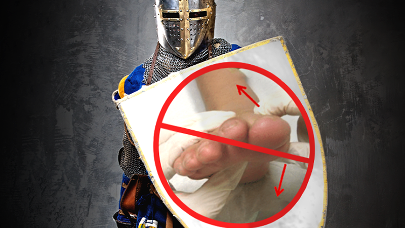
First, let’s recall what this horror of an examination the first ray excursion test actually is. Take a look at Figure 1. Recall this test is done by holding the foot in subtalar neutral with midtarsal joint locked. The forefoot is held with the contralateral hand, stabilizing it while the ipsilateral hand dorsiflexes and plantarflexes the 1st metatarsal head. Motion is estimated by comparing the residual dorsal and plantar position of the first metatarsal thumb in relation to the one holding the second metatarsal. Oh, thumbkin. You poor unwitting accessory to this criminal examination technique!

Now that we’ve identified our enemy, let’s find out why it’s a useless test. First, several studies have shown there is no highly accurate physical examination method to determine the actual amount of motion present, but let’s ignore that glaringly huge problem. Let’s also ignore that this is a static examination method that doesn’t correlate with gait.


The issue of focus for my crusade is the well-established reality that “first ray” dorsiflexion and plantarflexion is actually motion at multiple joints. Figure 2 combines outcomes from three important biomechanical kinematic studies.2,3,4 Each color in the image corresponds to a different study, and all three clearly show sagittal motion of the medial column is a combination of multiple joints. Additionally, the first metatarsal-medial cuneiform joint has much less motion than the naviculocuneiform joint. And I didn’t even include Phillips’ study using a 2D motion capture system that found “mild and inconsistent” motion of the medial cuneiform-first metatarsal joint with most of the plantarflexion motion of the first ray found to occur in the naviculocuneiform joint.1 If anything, the naviculocuneiform joint is our unsung hero of the medial column sagittal plane motion. Hear, hear NC joint!
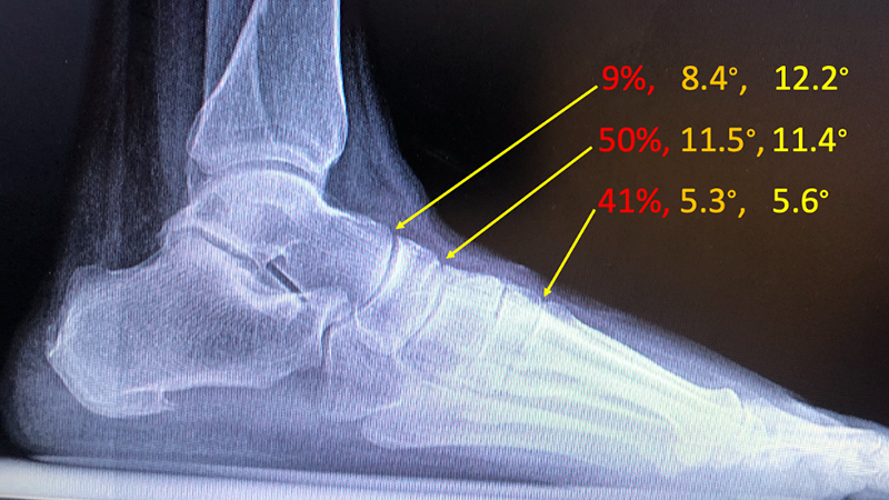
To summarize, these well-cited studies demonstrate a major percentage of medial column sagittal motion comes from the NC joint with less from the 1st metatarsal-medial cuneiform joint. “First ray” motion during the excursion test actually demonstrates combined motion from multiple joints.
“First ray” motion during the excursion test actually demonstrates combined motion from multiple joints.
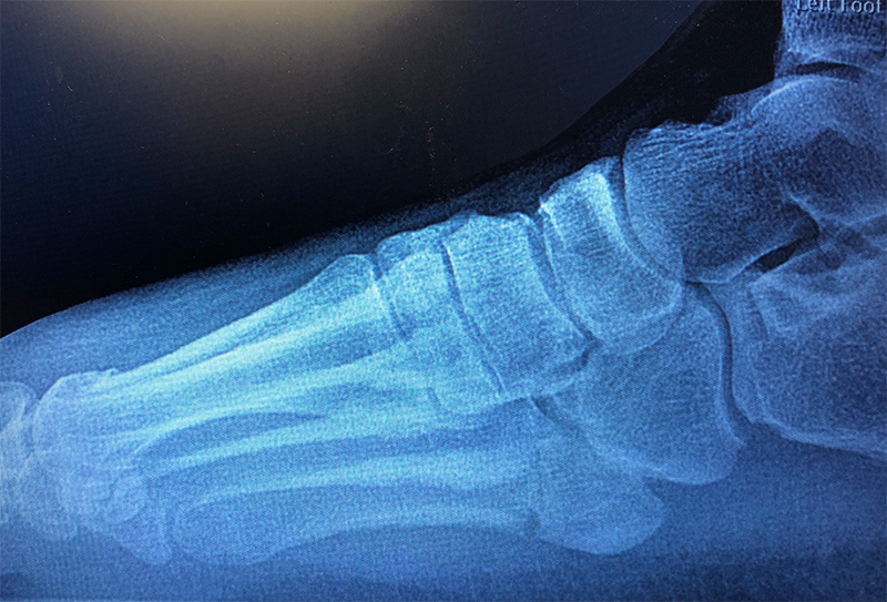
Figure 3 shows the radiographic manifestation of this fact.
What’s the consequence of this reality? The first ray excursion test is inaccurate and should not be used to determine first ray hypermobility, especially if one is using it to make surgical decisions.
The first ray excursion test is inaccurate and should not be used to determine first ray hypermobility, especially if one is using it to make surgical decisions.

If anything, this test simply examines overall medial arch and midfoot mobility. The first ray excursion test has had its day in the sun, but that’s over. Goodbye evil, inaccurate, and uninformative test. Burn in the fires of Mount Doom! Begone excursion test! Join your malicious siblings in Hades. Relegate the first ray excursion test to the dustbin of history, and you’ll be one step closer to biomechanical freedom.


-
Phillips RD, Law EA, Ward ED. Functional Motion of the Medial Column Joints of the Foot During Propulsion. J Am Podiatr Med Assoc. 1996 Oct;86(10):474-486.
Follow this link -
Roling BA, Christensen JC, Johnson CH. Biomechanics of the First Ray Part IV. The Effect of Selected Medial Column Arthrodeses. A Three-Dimensional Kinematic Study on a Cadaver Model. J Foot Ankle Surg. Sept-Oct 2002;41(5):278-285.
Follow this link -
Lundgren P, Nester C, Liu A, Arndt A, Jones R, Stacoff A, Wolf P, Lundberg A. Invasive in vivo measurement of rear-, mid- and forefoot motion during walking. Gait Posture. 2008 Jul;28(1):93-100.
Follow this link -
Nester CJ, Liu AM, Ward E, Howard D, Cocheba J, Derrick T, Patterson P. In vitro study of foot kinematics using a dynamic walking cadaver model. J Biomechan. 2007;40(9):1927-1937.
Follow this link
Major Sponsor


Comments
There are 0 comments for this article