| |
Robert Frykberg
DPM, MPH
PRESENT RI Editor
Diabetic Limb Salvage
|
Last time, we presented a case from the High Risk Clinic involving one of our regular diabetic neuropathic patients who required a left first ray amputation due to wet gangrene and presumed underlying osteomyelitis (November FootNotes). Since he had no signs to indicate peripheral arterial disease and his ankle-brachial indices and waveforms were normal, a vascular surgical consultation was unnecessary. He had healed his amputation site uneventfully over the course of four weeks and eventually resumed his usual activities (including riding his horses) wearing diabetic therapeutic footwear with custom insoles and his occasional cowboy boots. (After all, we live in the Wild West!).
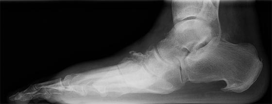
After several months, he noticed the insidious onset of painless swelling in the affected foot without any history of antecedent injury or recent open wounds. His foot X-rays are shown in Figures 1 and 2.
Figure 1. AP view of left foot |
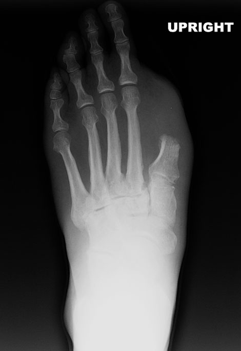 |
Figure 2. Lateral view of left foot |
 |
Most of you have already determined correctly the diagnosis for his current problem or have at least developed a list of differential diagnoses. Most obvious is the radiographic findings of the previous first ray amputation in Figure 1. There is no gas or defects in the soft tissues to indicate infection and the distal 1st metatarsal stump does not look to be abnormal, other than some minor periosteal proliferation. Nonetheless, let’s take a closer look at the magnified view in Figure 3.
Figure 3. Magnified AP view of left foot. |
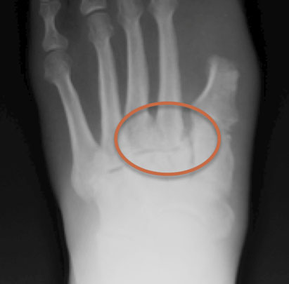 |
In any X-ray of a swollen neuropathic foot (with or without other changes ) one must look at the metatarsal bases and the Lisfranc joint. This is your key to making that very early diagnosis of Charcot arthropathy – as in this case. Within the red circle, we see that characteristic relatively subtle subluxation of the second metatarsal base at the articulation with its middle cuneiform (2nd tarsal-metatarsal joint). Even a one millimeter step–off is diagnostic for a Charcot foot in this setting. In fact, this is the first place I look when I am suspicious for Charcot, as I was with this patient. He also has an indistinct fracture of the lateral second metatarsal base and some fuzzy changes of the 3rd metatarsal base. In concert with the prior first ray amputation, this is a fairly common presentation for an acute Charcot foot. Looking again at Figure 2, we can see the midfoot collapse and the dorsal periosteal proliferation/fragmentation even involving the distal navicular. These changes confirm our diagnosis.
I have encountered this clinical scenario a number of times and it has even been described by Aragon–Sanchez and others. Amputation or partial amputation of the first ray results in a significant biomechanical insufficiency of the medial column. As I envision the pathomechanics, with the loss of a medial supporting structure, the force is shifted laterally to the lesser rays that are not as well equipped to withstand the forces. In those individuals with neuropathy predisposed (genetically) to develop Charcot arthropathy, even a minor injury or force can precipitate the initial dislocation or fracture that culminates in an acute Charcot foot. Therefore, it is prudent in such patients that we consider the possibility that neuropathic arthropathy can occur postoperatively and be vigilant in our follow-up care. While we are discussing this particular postoperative event, any foot surgery or injury might precipitate an acute event in the susceptible neuropathic patient. As always, your index of suspicion must always be high for this disorder.
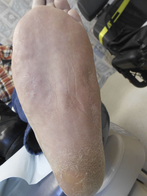
Once the diagnosis was made based on his clinical history, presentation and plain radiographs, the patient was first immobilized in a total contact cast (TCC) followed by a cast walker for approximately 12 weeks. We also provided him with a wheelchair to allow him to minimize walking on the injured foot. He subsequently converted to an inactive state, stabilized, and eventually resumed his protective weight-bearing with no recurrence of this problem, nor has he had any recurrent ulcers. (Figure 4)
Figure 4. Recent clinical photo of this patient�s foot. |
 |
Although it would have been optimal to make the diagnosis in Stage 0 before any radiographic changes became visible, we were fortunate to make a fairly early diagnosis during the active stage. Deformity was minimized and the patient was managed non-operatively with a good result. It bears repeating that a high index of suspicion is paramount in this regard – the earlier diagnosis can be made and treatment started, the more likely favorable results can be achieved.
Remember, if you look for Charcot you will find it. And the sooner, the better!
Until next time,

Robert Frykberg, DPM, MPH
PRESENT Editor
Diabetic Limb Salvage
Selected References:
- Frykberg RG, Zgonis T, Armstrong DG, Driver VR, Giurini JM, Kravitz SR, et al. Diabetic foot disorders. A clinical practice guideline (2006 revision). J Foot Ankle Surg. 2006;45(5 Suppl):S1-66.
- Aragon-Sanchez J, Lazaro-Martinez JL, Hernandez-Herrero MJ.
Triggering mechanisms of neuroarthropathy following conservative surgery for osteomyelitis. Diabet Med. 2010;27(7):844-847.
- Frykberg RG, Eneroth M. Principles of Conservative Management. In: Frykberg RG, ed.
The Diabetic Charcot Foot: Principles and Management. Brooklandsville, MD:
Data Trace Publishing Company; 2010:93-116.
- Boulton AJ, Armstrong DG, Albert SF, Frykberg RG, Hellman R, Kirkman MS, et al. Comprehensive foot examination and risk assessment: a report of the task force of the foot care interest group of the American Diabetes Association, with endorsement by the American Association of Clinical Endocrinologists. Diabetes Care. 2008;31(8):1679-1685.
- Rogers LC, Frykberg RG. The charcot foot. Med Clin North Am. 2013;97(5):847-856.
- Rogers LC, Frykberg RG, Armstrong DG, Boulton AJ, Edmonds M, Van GH, et al. The Charcot foot in diabetes. Diabetes Care. 2011;34(9):2123-2129.
- Jeffcoate WJ, Game FL. New Theories on the causes of the Charcot foot in diabetes.
In: Frykberg RG, ed. The Diabetic Charcot Foot: Principles and Management. Brooklandville, MD: Data Trace Publishing Company;2010:29-44.
|