| |
Robert Frykberg,
DPM, MPH
PRESENT Editor
Diabetic Limb Salvage |
Cases From the High Risk Foot Clinic
Neuroischemic Gangrene: Part 2
Last month we presented a case typical of those seen in High Risk Foot Clinics (refer to the June Foot Notes ezine). To recap, this patient was a 59 year old type 2 diabetic man with diabetes duration of 15 years and inadequately controlled with insulin (Hemoglobin A1c of 9.3%). He also had a history of severe coronary artery disease, Congestive Heart Failure, mild renal insufficiency (eGFR=50), peripheral neuropathy, and peripheral arterial disease (PAD). Of particular importance, he had already succumbed to a right below knee amputation (BKA) several years prior to his presentation to us. He was referred by his primary care physician with a diagnosis of gangrene and cellulitis of the left foot. (Figure 1).
Figure 1: The patient's left foot upon presentation |
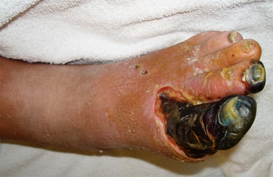 |
Briefly, our initial exam revealed an infected neuroischemic gangrene of the left great toe (and tip of second toe) with absent pedal pulses and an ABI of 0.65. There was no gas on plain Xrays. After culturing the wound, he was started empirically on vancomycin and piperacillin/tazobactam upon admission. Final wound cultures grew out only methicillin-sensitive staphylococcus aureus and his blood cultures were negative. The patient’s leukocytosis (12,500) rapidly resolved with antimicrobial therapy and his mild renal failure improved with hydration. But despite lack of fever, his erythema persisted.
After considering the patient’s presentation, examination, laboratory and diagnostic data, we posed five points to consider pertaining to his further management and care. For the sake of brevity, we will address them succinctly point by point. Recognize that there are many ways to approach any given patient according to currently accepted treatment guidelines. Nonetheless, the approach that my team took (as indicated below) may serve to guide you in the management of your own similar patients.
| 1. What consultations are required?
We typically consult our Medical service for all such patients to assist in assessing and managing not just the diabetes, but other comorbidities as well (Cardiac, Hypertension, renal insufficiency, etc.). If renal function deteriorated, we would have also consulted Nephrology. In a patient such as this where we have already diagnosed ischemia on the basis of our clinical examination and vascular lab studies, it is critical to obtain a Vascular Surgery (or vascular interventionalist) consult to evaluate revascularization options to restore perfusion to the ischemic extremity. Although we did not need to do so in this case, an infectious disease consultation is frequently advised, especially for resistant infections.
Furthermore, we routinely ask for a Nutrition consult to assist in managing the patients’ diet and caloric requirements during such stressful periods.
2. Is antimicrobial therapy appropriate?
In this case, the empirical antimicrobial coverage was overly broad once the final culture results were obtained. Clinically, the patient was improving but we did not need to cover for MRSA or gram negative bacilli. At this juncture is appropriate to narrow the antimicrobial regimen to cover the isolate returned: methicillin sensitive staphylococcus aureus (MSSA). We chose a first generation cephalosporin (cefazolin), but one could just as well prescribe oxacillin, nafcillin, or clindamycin in many cases depending upon sensitivities and allergy profiles. Granted, the anaerobic cultures take a week or so to return, but we felt safe in this gap in coverage for this patient once his clinical signs improved.
3. Are there other studies that we specifically need to order?
Since osteomyelitis (or extent of osteomyelitis) is always a concern in such patients, we routinely would order an MRI or PET/CT scan. Alternatively, a three phase bone scan combined with a leukocyte scan would be able to identify foci suspicious for osteomyelitis. Most pressing, however, would be the need for an angiogram in anticipation of revascularization. This is critical and we usually leave this decision up to our vascular surgeons (who might also perform the study as well). If concerns arise pertaining to cardiac function (in anticipation of likely surgical intervention), our Medical team would consult Cardiology and order Cardiac perfusion stress test and/or cardiac echo studies.
4. What surgical procedures would you anticipate this patient requiring?
Since the patient has a gangrenous forefoot, we know that amputation will be required. The ultimate questions remains at which level could healing be predicted? With poor perfusion to the foot the likelihood of healing a primary partial foot amputation is low. Since no urgent open amputation was necessary to control infection, we chose to await vascular intervention prior to proceeding with our surgical intervention.
5. Does he need another BKA to resolve his problem?
Our ultimate goal is always limb salvage whenever feasible and in most cases major amputation can be avoided. In a patient such as this who has already lost the contralateral limb, it becomes even more important. Revascularization, either by endovascular or open surgical procedure, is the key to sparing the limb. Once perfusion to the foot can be restored, a limb salvaging (and foot sparing) partial foot amputation can be performed where tissue loss is confined to the forefoot. |
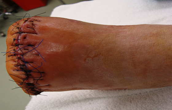
So how did we proceed with this patient? After revascularization with a femoral- tibial bypass graft we were able to subsequently perform a transmetatarsal amputation with primary closure. (Figure 2)
Figure 2: TMA immediately postoperative. Primary closure is always sought in such cases when no active infection exists at the amputation margins |
 |
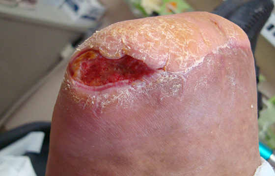
One month or so postoperatively, the patent developed a partial dehiscence of his incision. (Figure 3)
Figure 3: Partial dehiscence of incision occurred 5 weeks after surgery, but no active infection is noted. This was treated with human fibroblast derived dermal substitutes |
 |
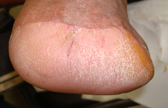
Since it was not infected, we were able to treat in the clinic with frequent applications of human fibroblast derived dermal substitute to generate healthy granulations and tissue regeneration. Although these patients often have a prolonged healing time when pulsatile flow is not restored to the foot (as in this case), the patient did eventually heal his amputation. (FIGURE 4)
Figure 4: Healed incision 5 months postoperatively |
 |
Once healed he was dispensed diabetic therapeutic footwear with custom insoles and is followed every three months for ongoing prevention visits.
I’m sure that you’ll agree that this is a fairly common clinical scenario for many of our patients. By using established guidelines for management and careful assessment of the patient (and wounds), you can be very successful in your limb salvage efforts. As exemplified in this case, multidisciplinary management often makes the difference between success and failure.
We welcome your opinions, concerns, and suggestions. If you have an interesting case or a troubling circumstance that you would like to share with fellow PRESENT Diabetes members, please feel free to comment on eTalk.
Until next time,,

Robert Frykberg, DPM, MPH
PRESENT Editor
Diabetic Limb Salvage
References:
-
Lipsky BA, Berendt AR, Cornia PB, et al. 2012 Infectious Diseases Society of America
clinical practice guideline for the diagnosis and treatment of diabetic foot infections.
Clin Infect Dis. Jun 2012;54(12):e132-173.
-
Frykberg RG, Zgonis T, Armstrong DG, et al. Diabetic foot disorders. A clinical practice guideline (2006 revision). J Foot Ankle Surg 2006;45:S1-66.
-
Aragon-Sanchez J. Seminar review: A review of the basis of surgical treatment of diabetic foot infections. Int J Low Extrem Wounds 2011;10:33-65.
-
Boulton AJ, Kirsner RS, Vileikyte L. Clinical practice. Neuropathic diabetic foot ulcers.
N Engl J Med 2004;351:48-55.
- Lepantalo M, Apelqvist J, Setacci C, et al. Chapter V: Diabetic foot. Eur J Vasc Endovasc Surg 2011;42 Suppl 2:S60-74.
|