| |
Robert Frykberg,
DPM, MPH
PRESENT Editor,
Diabetic Limb Salvage |
Case Study: Diabetic Foot Flu, Part 2
– Cases from the High Risk Foot Clinic
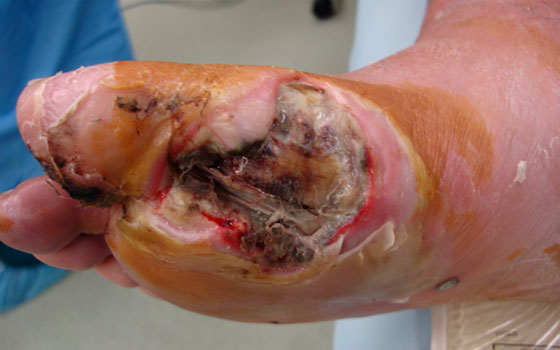
Last month, we presented the first in a series of Cases from the High Risk Foot Clinic in which we described that typical Friday afternoon diabetic patient presenting to the Emergency Room; the kind with an indolent neuropathic foot ulcer who had developed flu-like symptoms over the preceding week. I term this illness the "Diabetic Foot Flu". We have learned from experience that any diabetic patient with neuropathy and a painless foot ulcer who develops the onset of pain in the previously insensitive foot must be suspected of having an acute infection until proven otherwise. This is simply a good premise to manage your patients by. As was the case with our patient, he developed an ascending necrotizing infection (necrotizing fasciitis) emanating from the chronic wound under his first metatarsal. (Figure 1). He was quite sick, with abnormal electrolytes, blood gases, hypotension, hyperglycemia, leukocytosis, septicemia (Beta hemolytic streptococci), and high fever (see last month’s issue for particulars). Evaluation of his foot revealed a foul smelling wound with tendon and bone exposed, crepitus on motion of the great toe, ascending cellulitis, and purulent drainage. Critically important, we noted the presence of ascending gas in his foot and lower leg. This, as always, represented a surgical emergency and he was taken to the operating room within two hours.
Figure 1: Initial presentation |
 |
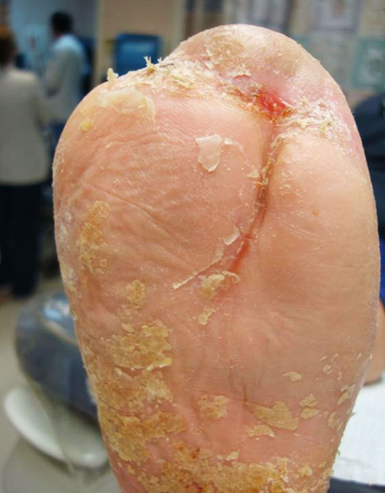
Our initial procedure, of course, was drainage and control of infection with an open first ray amputation as well as wide incision and drainage (I&D) with debridement of his lower leg. (Figure 2). Concurrently, he had both an intravenous line placed for fluid resuscitation, intravenous medications (including insulin drip), and antibiotics; as well as an arterial line for better monitoring of his hemodynamic status. Empirical broad spectrum antimicrobial therapy was initiated with Vancomycin and Piperacillin/tazobactam to cover most potential Gram positive pathogens (including Methicillin-resistant staphylococcus aureus [MRSA]), anaerobes, and Gram negative bacilli. His wounds were packed open to allow for drainage and subsequent inspection. He went to the Intensive care unit for close monitoring by our medical team postoperatively. His condition improved overnight, but areas of necrosis were still noted on the skin edges and at the base of both the leg and foot wounds several days later. (Figure 3) Blood and wound cultures returned growth only of beta hemolytic streptococci; his antibiotic coverage was then narrowed to Penicillin 4 million units every four hours by our Infectious Disease specialist. As is frequently necessary in such cases, he was subsequently taken back to the operating room several times for further debridement of his wounds, including revision to an open transmetatarsal amputation (TMA). (Figure 4) The size and nature of his wounds also necessitated using negative pressure wound therapy (NPWT) to assist in managing drainage and the wound beds themselves. (Figure 5.) Luckily, the patient was not ischemic and had easily palpable pedal pulses. He therefore responded fairly rapidly with the development of healthy granulations. We expedited wound closure thereafter with a combination of therapies including NPWT, pulsed radio frequency energy, and a human fibroblast derived dermal substitute (HFDS) over the course of the next several months. Most of his treatment with these advanced therapies took place in the outpatient setting, with weekly clinic visits and home health care visits to change the NPWT dressings. Although he returned to the hospital once in the ensuing months for a tendo-Achilles lengthening and split thickness skin graft to a small wound on the dorsum of his foot, he was completely healed approximately 8 months after initial presentation. (Figures 6 and 7). He is now followed regularly in our Podiatry (Prevention) Clinic and wears prescribed therapeutic footwear with custom insoles.
Figure 2 |
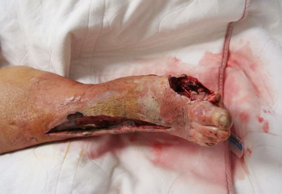 |
Figure 3 |
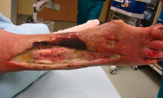 |
Figure 4 |
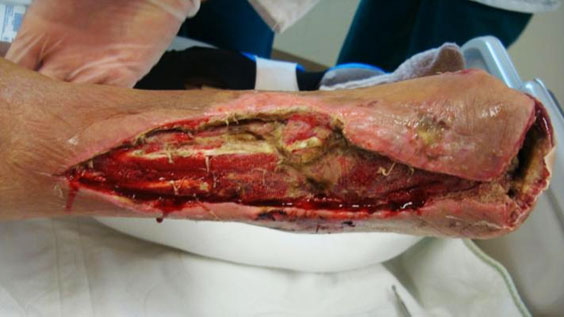 |
Figure 5 |
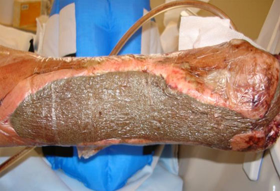 |
Figure 6 |
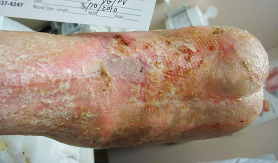 |
Figure 7 |
 |
This brief case illustrates the challenge of dealing with acute necrotizing infections in diabetic patients without having to resort to immediate major amputation. Although every facet of care was not discussed in this regard, a successful outcome was achieved through following basic principles of care for such limb-threatening problems: early and aggressive surgical control of infection, culture guided antimicrobial therapy, wound management based upon appearance and progress of healing, rational use of advanced therapies, and multidisciplinary management. Following these basic steps for any patient presenting with acute infections will result in better outcomes for most patients.
We welcome your opinions, concerns, and suggestions. If you have an interesting case or a troubling circumstance that you would like to share with fellow PRESENT Diabetes members, please feel free to comment on eTalk.
eTalk for this ezine?

Best regards,

Robert Frykberg, DPM, MPH
PRESENT Editor,
Diabetic Limb Salvage
References:
- Lipsky BA, Berendt AR, Cornia PB, et al.: 2012 Infectious Diseases Society of America clinical practice guideline for the diagnosis and treatment of diabetic foot infections. Clin Infect Dis 2012; 54(12): e132-73.
- Richard J-L, Sotto A, Lavigne J-P: New Insights in Diabetic Foot Infection. World Journal Diabetes.(2011), 2:24-32
- Frykberg RG, Zgonis T, Armstrong DG, et al.: Diabetic foot disorders: a clinical practice guideline (2006 Revision). . J Foot Ankle Surg 2006; 45(5 Suppl): S1-66.
|